User talk:Ancheta Wis/c
The following C language fragment is syntactically correct, but performs an operation that is not semantically defined when p is a null pointer; the operations p->real and p->im would have no meaning unless *p were initialized to some hardware memory address, rather than NULL. In practice, the second line would have to be wrapped by a conditional statement to prevent execution when *p is NULL. Even then, the code fragment would fail a code review:
complex *p = NULL;
complex abs_p = sqrt (p->real * p->real + p->im * p->im);
Bib listing for Memory-prediction framework
C[edit]
The visual system is what allows us to see. It consists of:
- The eye, especially the retina
- The optic nerve
- The optic chiasm
- The optic tract
- The lateral geniculate nucleus
- The optic radiations
- The visual cortex
The task of the visual system is to interpret what would otherwise be a two-dimensional image of the world as a moving, colored three-dimensional world.
This article mostly describes the visual system of mammals, although other "higher" animals have similar visual systems.
 Light is inverted by the lens and projected onto the retina; blue-attuned cone cells will be most strongly stimulated by blue light, while yellow/red-attuned cone cells will not be. |
Eye[edit]
The eye is a complex biological device, considered by some to be miraculous. The eye functions remarkably like a CCD camera, taking visible light and converting it into a stream of information that can be transmitted via nerves.
Light enters the eye, passing through the cornea and the pupil (controlled by the iris) and is refracted by the lens. The lens inverts the light and projects an image onto the retina.
The retina consists of a large number of photoreceptor cells which contain a particular protein molecule: the photopigment called rhodopsin. When rhodopsin is struck by a photon (a particle of light) it transmits a signal to the cell; the more photons strike the cell, the stronger the signal will be. In some animals, like humans, cone cells contain cone opsin molecules attuned to specific wavelengths of light; i.e., a blue cone cell contains opsin most attuned to blue-wavelength light and will most strongly be stimulated by blue-wavelength light, while a yellow-red cone cell will only be weakly stimulated by blue-wavelength light. This gives the ability to distinguish color.
Optic nerve[edit]
Following some rudimentary processing (mostly involving color boundaries), the information about the image received by the eye is transmitted to the brain via the optic nerve. In humans, the optic nerve is the only sensory system that is connected directly to the brain and does not connect through the medulla, due to the necessity of processing the complex visual information quickly.
Optic chiasm[edit]
The optic nerves from both eyes meet and cross at the optic chiasm, at the base of the frontal lobe of the brain. At this point the information from both eyes is combined and split according to the field of view. The corresponding halves of the field of view (right and left) are sent to the left and right halves of the brain, respectively (the brain is cross-wired), to be processed. That is, though we might expect the right brain to be responsible for the image from the left eye, and the left brain for the image from the right eye, in fact, the right brain deals with the left half of the field of view, and similarly for the left brain. (Note that the right eye actually perceives part of the left field of view, and vice versa).
Optic tract[edit]

Information from the right visual field (now on the left side of the brain) travels in the left optic tract. Information from the left visual field travels in the right optic tract. Each optic tract terminates in the lateral geniculate nucleus (LGN) in the thalamus.
Lateral geniculate nucleus[edit]
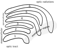
The lateral geniculate nucleus (LGN) is a sensory relay nucleus in the thalamus of the brain. The LGN consists of six layers. In humans and some other primates such as macaques, layers 1, 4, and 6 correspond to information from one eye; layers 2, 3, and 5 correspond to information from the other eye. Layer one (1) contains M cells, which correspond to the M cells of the optic nerve of the opposite eye. Layers four and six (4 & 6) of the LGN also connect to the opposite eye, but to the P cells of the optic nerve. By contrast, layers two, three and five (2, 3, & 5) of the LGN connect to the M cells and P cells of the optic nerve for the same side of the brain as its respective LGN. The six layers of the LGN are the area of a credit card, but about three times the thickness of a credit card, rolled up into two ellipsoids about the size and shape of two small bir. The neurons of the LGN then relay the visual image to the primary visual cortex (V1) which is located at the back of the brain (caudal end).
Optic radiations[edit]
The optic radiations carry information from the midbrain lateral geniculate nucleus to layer 4 of the visual cortex. The P layer neurons of the LGN relay to V1 layer 4C β. The M layer neurons relay to V1 layer 4C α. At this juncture, the image path ceases to be straightforward; there is more cross-connection within the visual cortex.
Visual cortex[edit]
The visual cortex is the most massive system in the human brain and is responsible for higher-level processing of the visual image. It lies at the rear of the brain (highlighted in the image), above the cerebellum. The interconnections between layers of the cortex, the thalamus, the cerebellum, the hippocampus and the remainder of the areas of the brain are under active investigation. Currently, much of our knowledge stems from patients with damage to known areas of the brain, with a corresponding study of the cognitive functions which have been spared.
- Memory-prediction framework
- Martin J. Tovée (1996), An introduction to the visual system. Cambridge University Press, ISBN 0521483395 (References, pp.180-198. Index, pp.199-202. 202 pages.)
- David Marr (1982), Vision: A Computational Investigation into the Human Representation and Processing of Visual Information. San Francisco: W. H. Freeman.
- David H. Hubel (1989), Eye, Brain and Vision. New York: Scientific American Library.
- Torsten Wiesel and David H. Hubel
- Matthew Schmolesky, The Primary Visual Cortex
- R.W. Rodiek (1988). "The Primate Retina". Comparative Primate Biology Vol. 4 of Neurosciences. (H.D. Steklis and J. Erwin, editors.) pp. 203-278. New York: A.R. Liss.

Crick p 87 Patricia Churchland, neurophilosopher [http://www.auto.ucl.ac.be/EYELAB/neurophysio/light
